CVC Insertion SimulatorⅡ <M93UB>

| ⇨Products |
CVC Insertion Simulator Ⅱ
CVC Insertion Simulator2 offers efficient training in both landmark and ultrasound-guided central venous catheterization. For both procedures, introductory training is prepareso that trainees can develop their skills in sequence. This is one of a kind simulator that provides simulation of common complications including mislodging.
Features
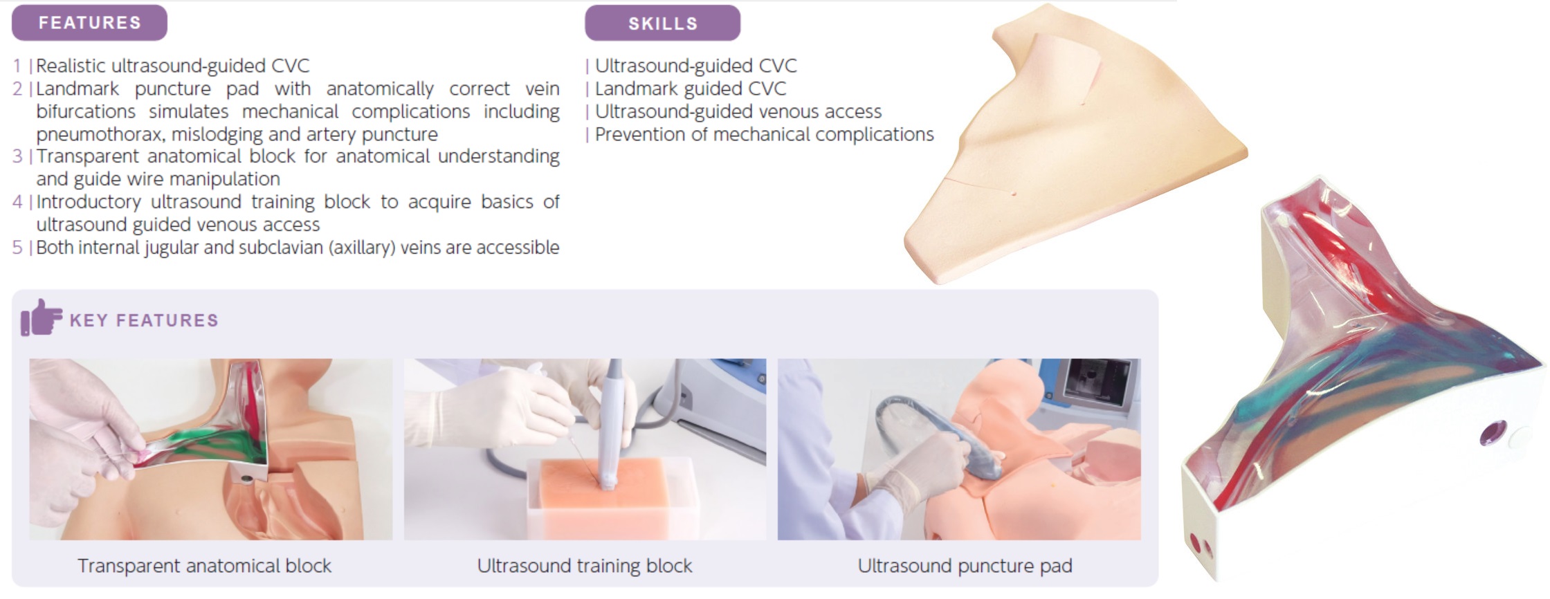
- This simulator includes three interchangeable pads for the puncture area, landmark puncture pad, ultrasound puncture pad and transparent cannulation block.
- The trainee will learn three skills: how to make a safer puncture, how to avoid possible complications that may accompany the puncture, how to insert the catheter into the correct position. When the procedure is performed incorrectly, the error will show immediately by feedback.
- Each training pad, placed at the right upper breast and right half of the neck covering the puncturing sites and catheter routes is a precise, life-size model incorporating the anatomical structure of bones, veins, arteries and upper lung.
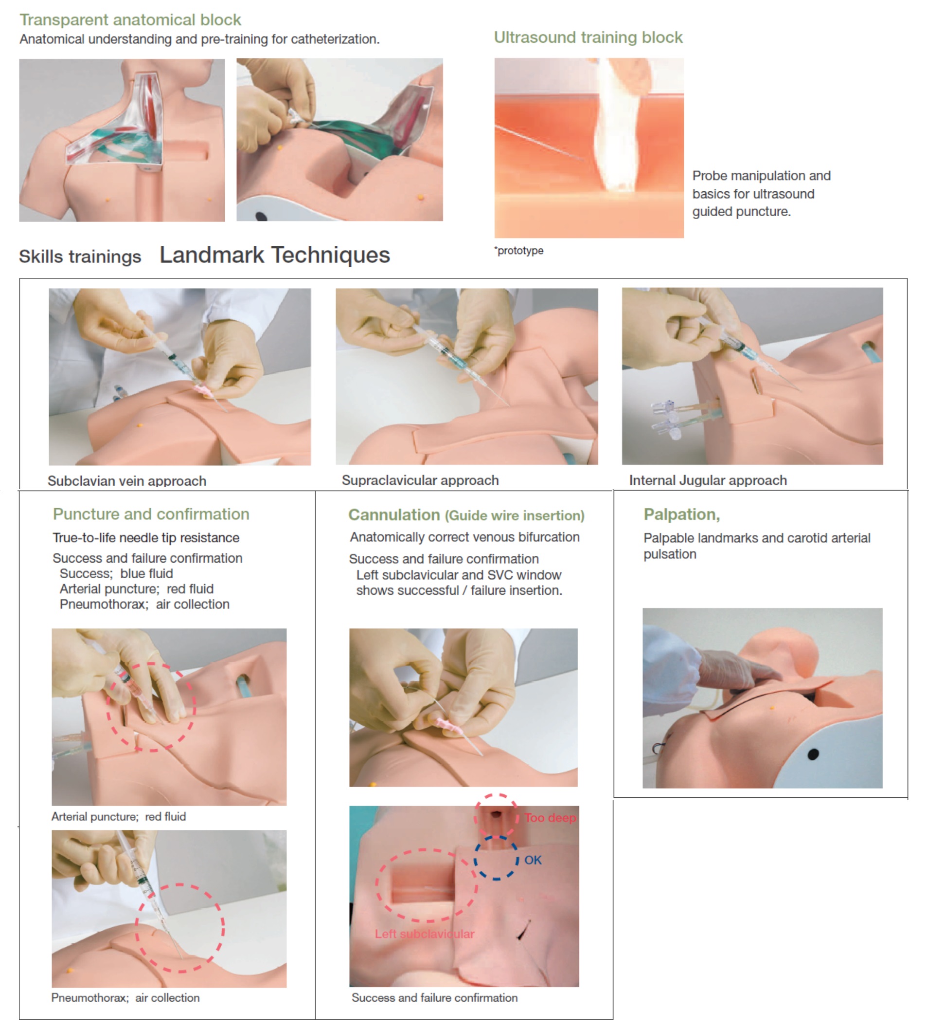
- The landmark puncture pad is designed to provide training in safely puncturing the subclavian vein or internal jugular vein, and inserting a catheter into SVC. Carotid artery pulsation is palpable.
- The ultrasound puncture pad allows training in internal jugular vein puncturing under ultrasound scanning. Clear scanning image facilitates understanding of how to distinguish vein from artery and perform safe puncture while watching an ultrasound monitor.
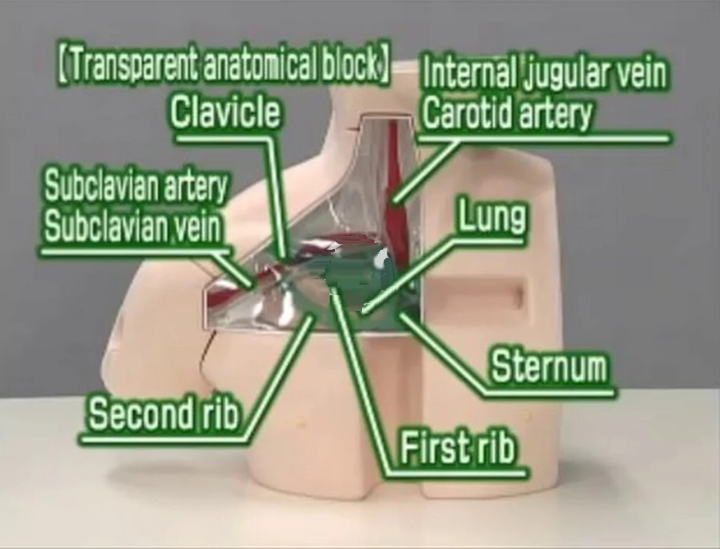
- A transparent block for three-dimensional anatomical understanding also works as an effective training tool for developing guide wire insertion skills.
Production Supervision:
Akira Okada, M.D., F.A.C.S., President
Osaka Medical Center and Research Institute for Maternal and Child Health
Masahiro Tanabe, M.D., Ph.D., Director
Postgraduate and Continuing Medical Education Center Chiba University School of Medicine
Kinya Sando, M.D., Ph.D., Professor
Division of Human Dietetics
Graduate School of Human Science Osaka Shoin Women's University
Masanori Hoki, M.D., Ph.D.
Head of Nutrition Management Center, Head of Community Health Service Center
Chief Pediatric Surgeon
Division of Pediatric Surgery Rinku General Medical Center Izumisano Municipal Hospital
Skills & Training
Three CVC approaches with landmark method:
- subclavian vein approach (a),
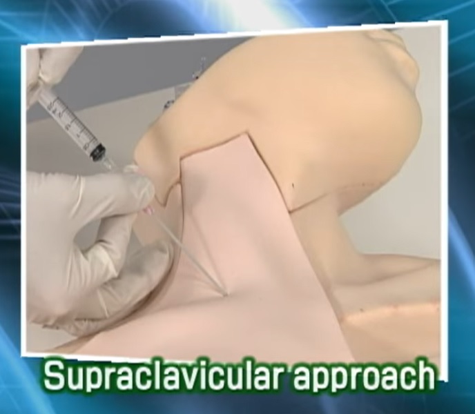
- supraclavicular approach (b) and
- internal jugular vein approach (c).
Ultrasound guided CVC from internal jugular vein and axillary approach.
Thorough procedure from puncture to cannulation
Anatomical learning
Complications indications:
- Artery puncture
- Pneumothorax
- Mislodging/malposition

Specifications
Set includes:
- 1 male upper torso manikin
- 1 landmark puncture pad
- 1 ultrasound puncture pad
- 1 transparent anatomical block
- 1 introductory ultrasound training block
- 1 skin for cannulation training
- 1 red coloring power
- 1 blue coloring power
- 1 air bulb
- 2 plastic jars
- 1 irrigation bottle
- 1 syringe
- 1 sample needle
Manikin size:
W40xD20xH34 cm, 2.2 kg
Packing size:
52 x 46 x 39 H cm, 9 kg
*Specifications are subject to change.
| ⇨Products |