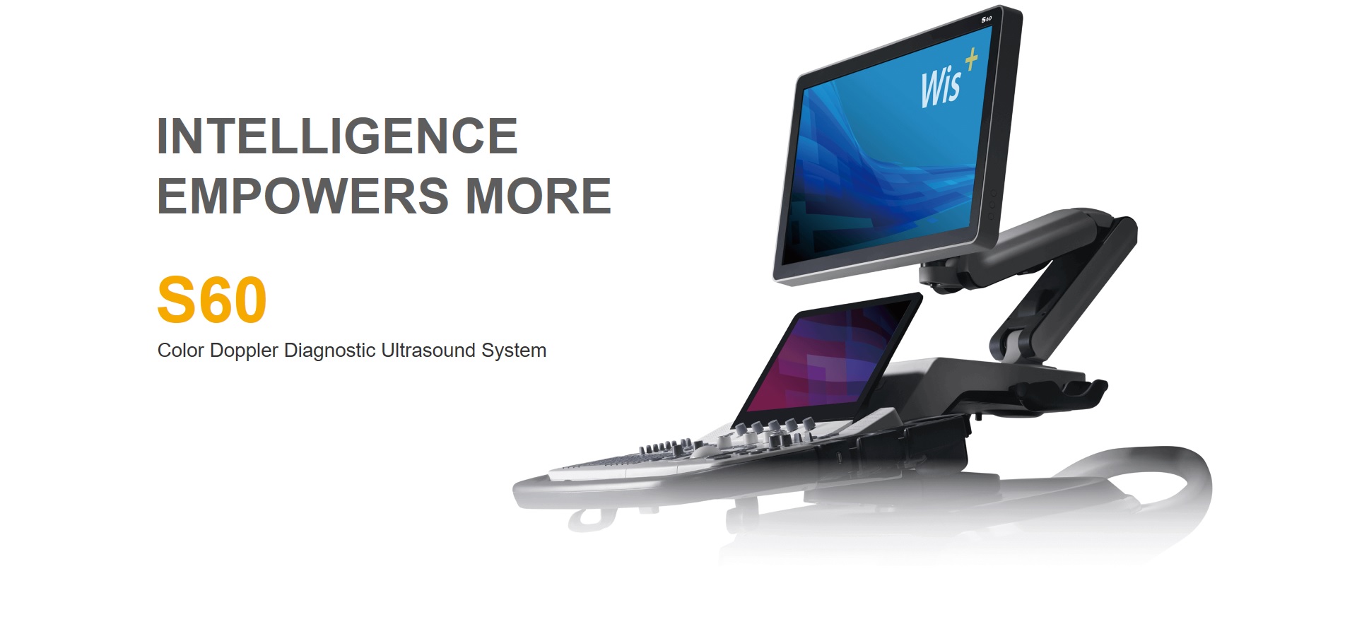
SonoScape S60
| ⇦ | ⇩ S60 Images ⇩ | Home ⊳ |
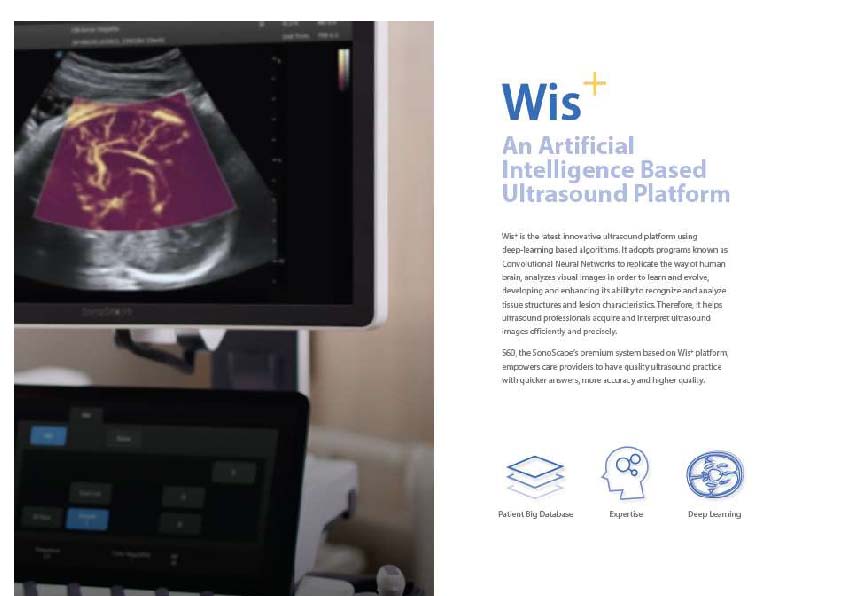
and interpret ultrasound images more efficiently and precisely.
It is a premium system that keeps your practice at the forefront of ultrasound examinations and
enables you to provide meticulous care for patients.
| ⇦ | ⇩ More Product information ⇩ | Home ⊳ |
| ⇦ | ⇩ More Product information ⇩ | Home ⊳ |
Wis+
Artificial Intelligence Based Ultrasound PlatformWis+ uses deep-learning algorithms to learn and evolve like human brain.
With this intelligent platform designed to actively adapt and analyze images,
exams are easy to perform and a new level of workflow is achieve.
| ⇦ | ⇩ More Product information ⇩ | Home ⊳ |
| ⇦ | ⇩ More Product information ⇩ | Home ⊳ |
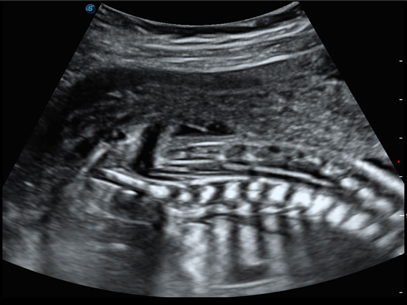
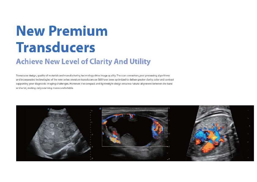
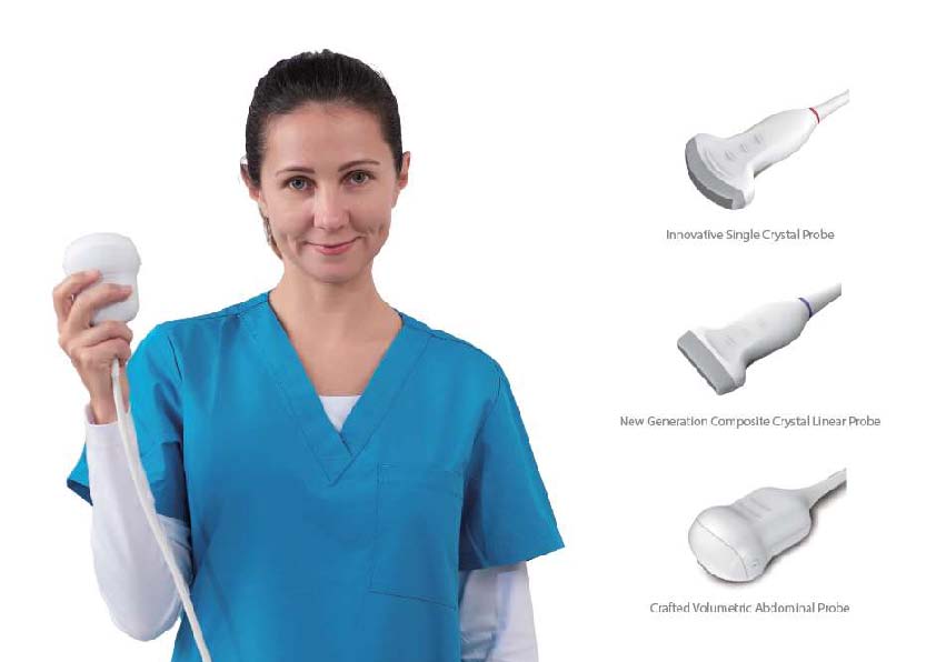
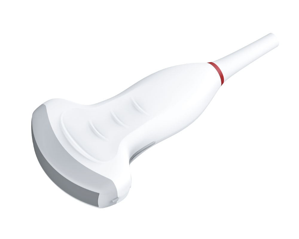
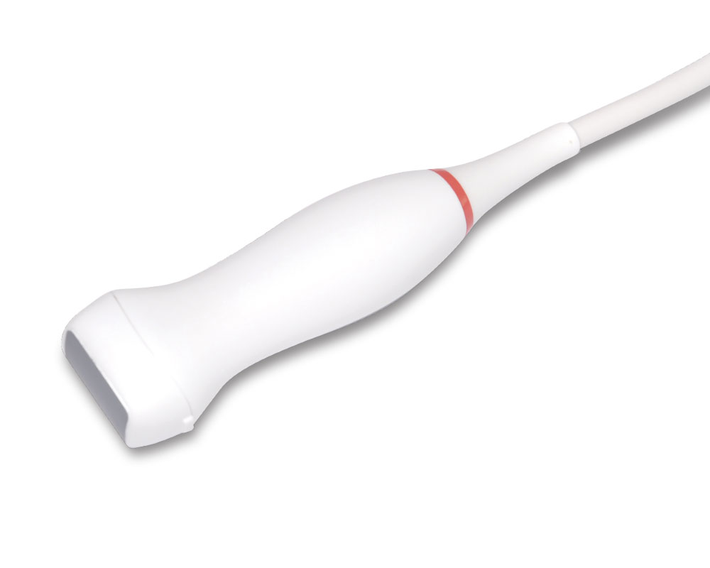
New Premium TransducersNew Level of Clarity and UtilityThe scan converters and post processing of the |
 |
|
 |
 |
 |
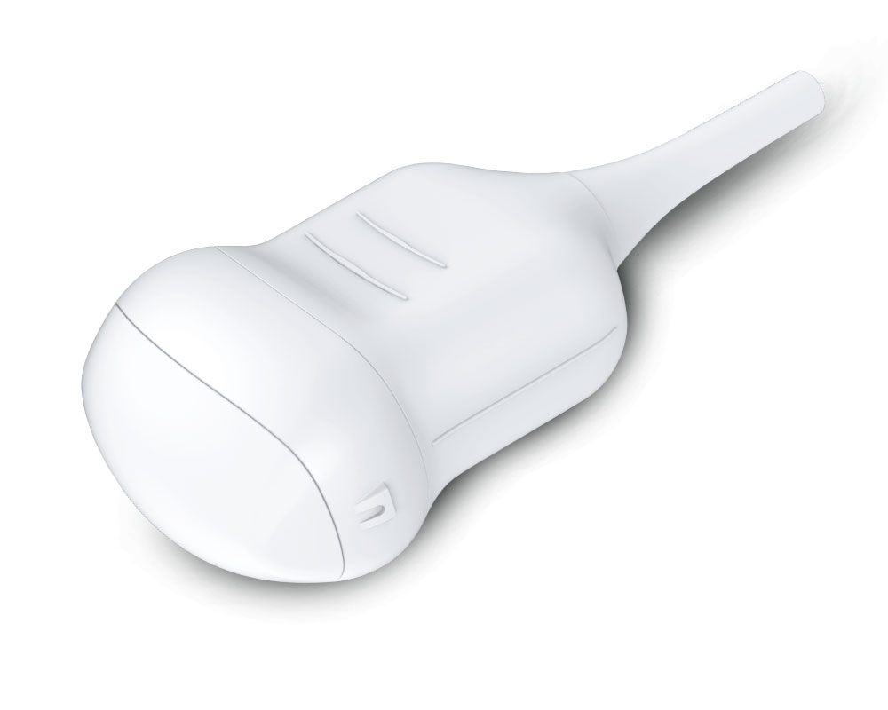 |
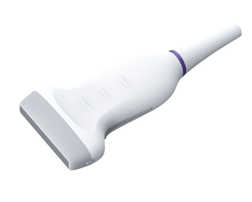
| New Generation Composite Crystal Linear Array |
Innovative Single Crystal Convex Array |
Innovative Single Crystal Phased Array |
Crafted Volumetric Abdominal Probe |
| ⇦ | ⇩ More Product information ⇩ | Home ⊳ |
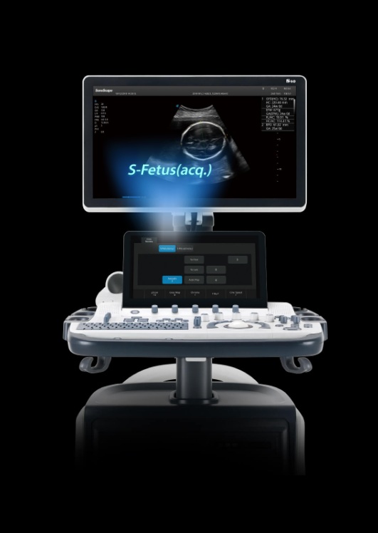
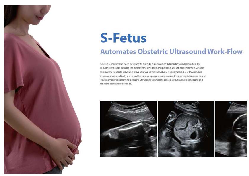
S-Fetus
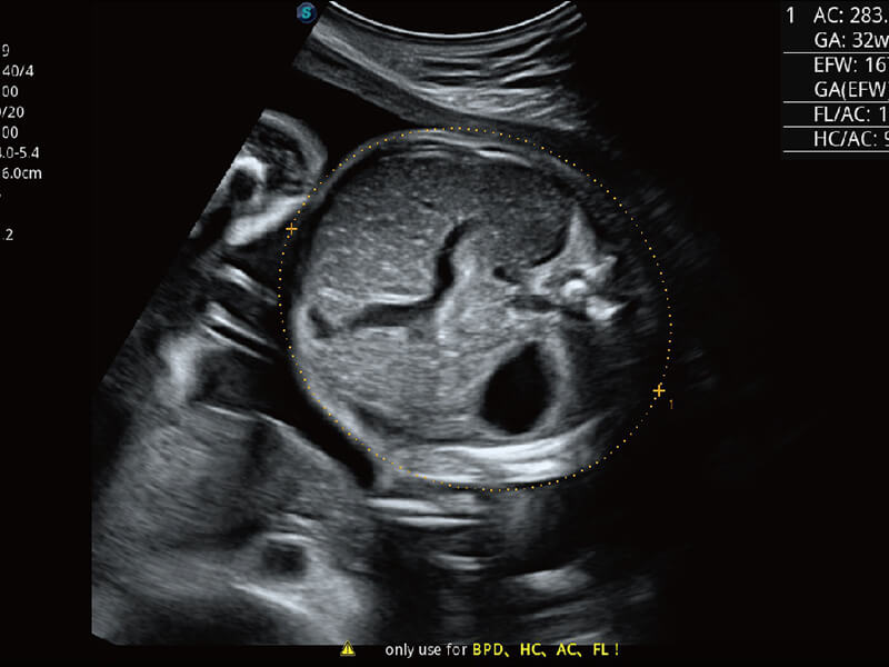
Automates Obstetric Ultrasound Work-Flow| Acquisition of fully automatic Fetal Biometries (Available with BPD / AC / HC / FL / AFI / PL) Automatic measurements and reports |
|
 |
|
| 80%fewer keystrokes 90%less time More than95%accuracy |
|
S-Fetus is a function that simplifies the standard obstetric ultrasound procedure.
With one touch, it selects the best section image and automatically performs the various measurements required to monitor fetus growth and development,
transforming obstetric ultrasound exams into an easier, faster, more consistent and far more accurate experience.
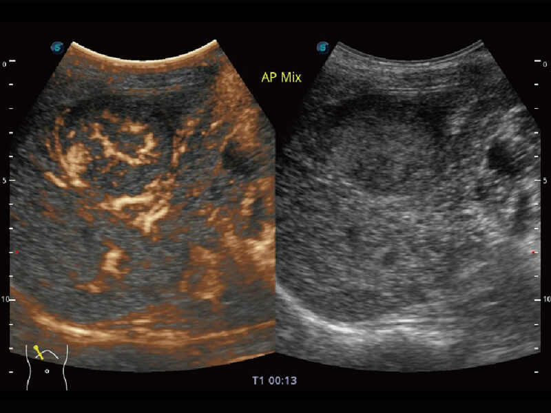
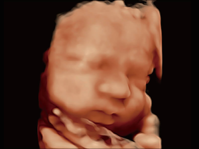
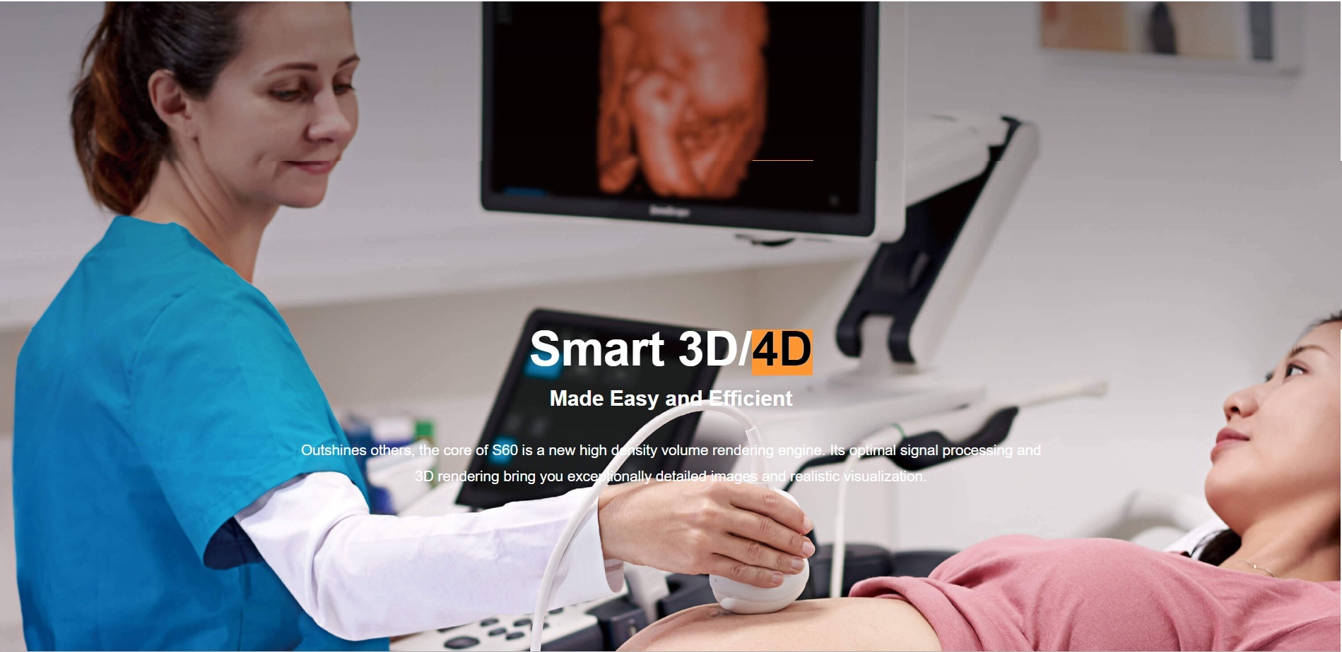
Smart 3D/4D
| Outshines others, the core of S60 is a new high density volume rendering engine. Its optimal signal processing and 3D rendering bring you exceptionally detailed images and realistic visualization. | ||
 |
||
|
|
|
|
|
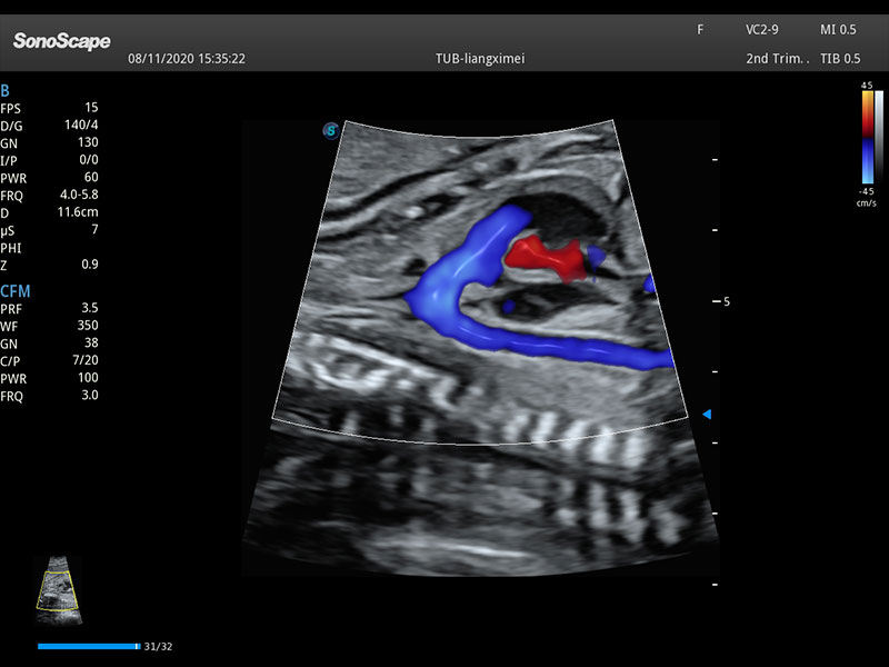
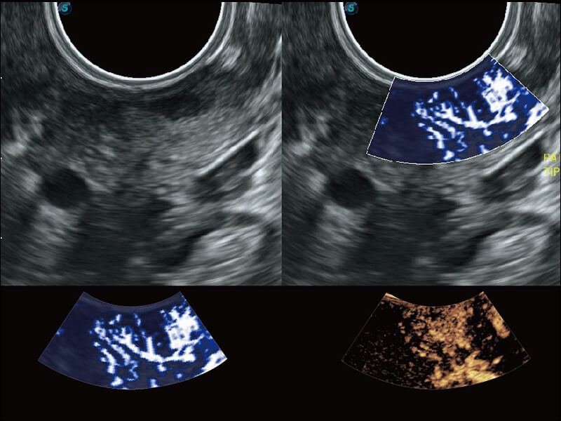
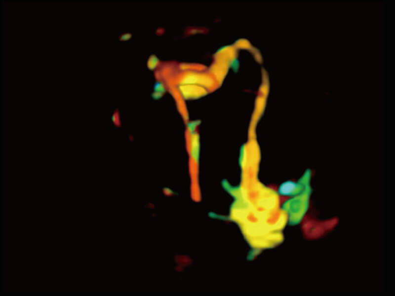
Fallopian Tube with CEUS |
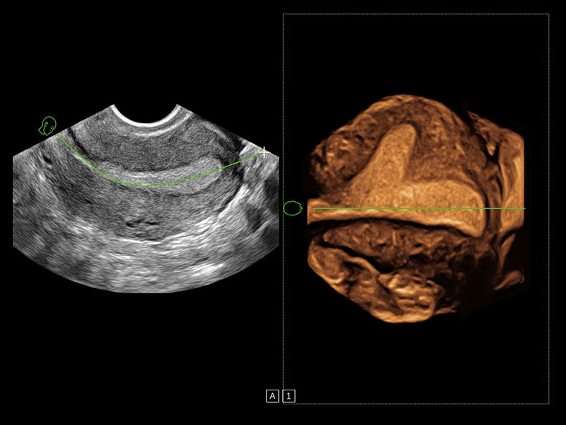
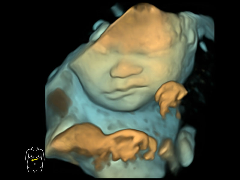
Fetal Face with S-Live |
Fetus Face |
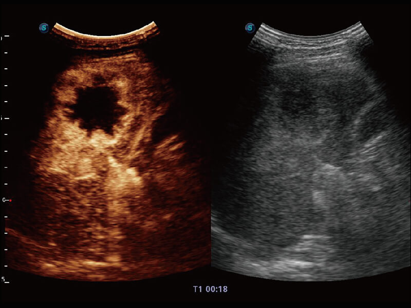
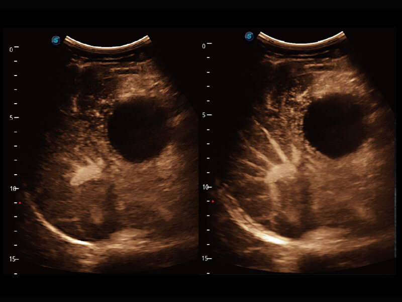
S-Live
Applies a virtual light source and advanced rendering technology that simulates the realistic skin effect.
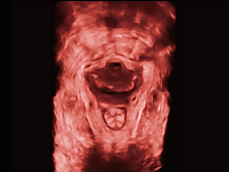
S-Live Silouette
It allows to observe the internal anatomical structure of any volume previously acquired automatically.
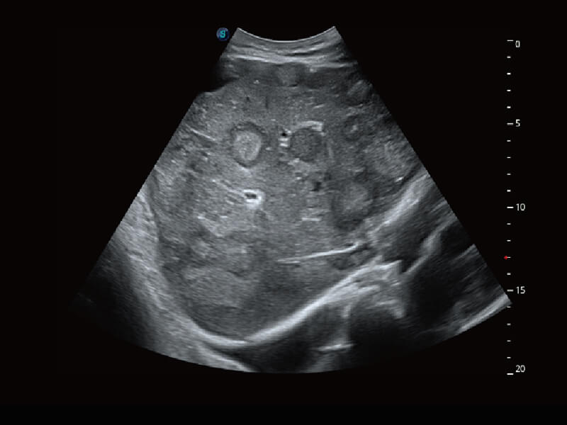
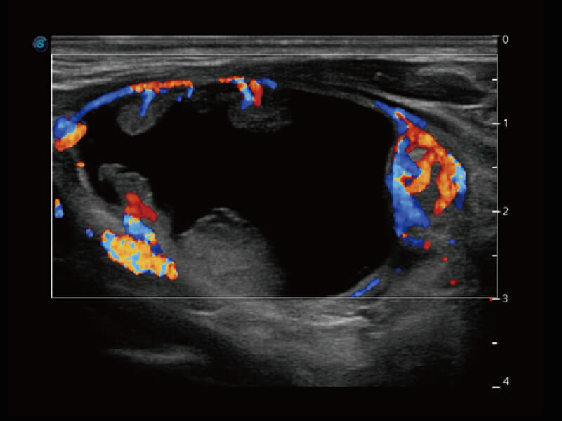
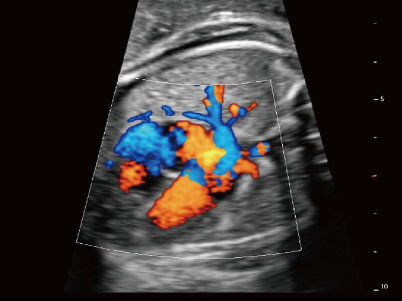
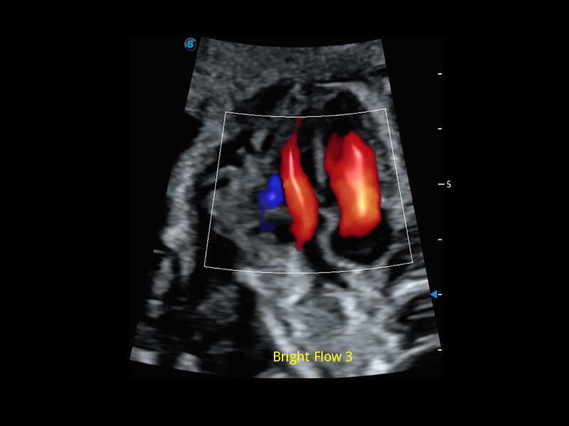
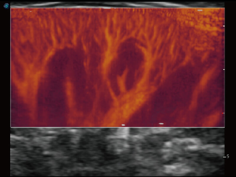
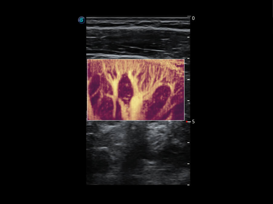
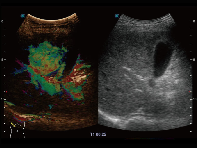
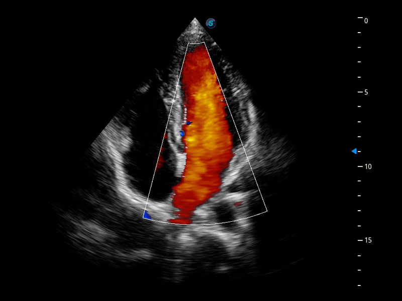
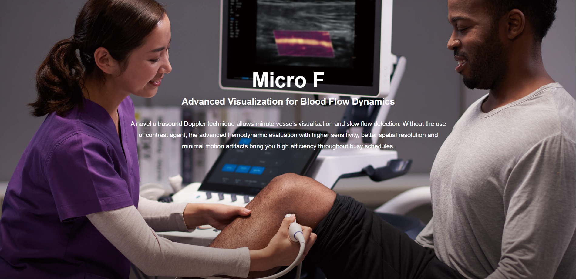
Micro F
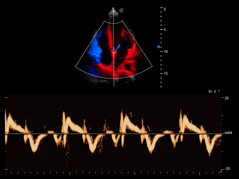
The adaptive Matrix E filter effectively distinguishes the blood flow of individual tissues, artifacts and flows at low speed
The algorithm helps to visualize the low blood flow in a stable and continuous way
Advanced Visualization for Blood Flow Dynamics
A novel ultrasound Doppler technique allows minute vessels visualization and slow flow detection. Without the use of contrast agent, the advanced hemodynamic evaluation with higher sensitivity, better spatial resolution and minimal motion artifacts bring you high efficiency throughout busy schedules.
⇦ ⇧ Top ⇧ Home ⊳