LM-099
Ultrasonic Bronchoscopy Simulator LM-099


Outline
This model can be used not only for ultrathin bronchoscope insertion training, but also for endobronchial ultrasound-guided transbronchial needle aspiration (EBUS-TBNA) training.
Features
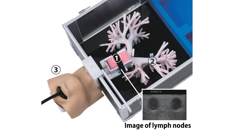
- Visualizing ultrasonic bronchoscopy images of lymph nodes embedded in the puncture site enables highly realistic definitive diagnosis of cancer metastases to the hilar and mediastinal lymph nodes, as well as practical training for puncturing the target lymph node. ①
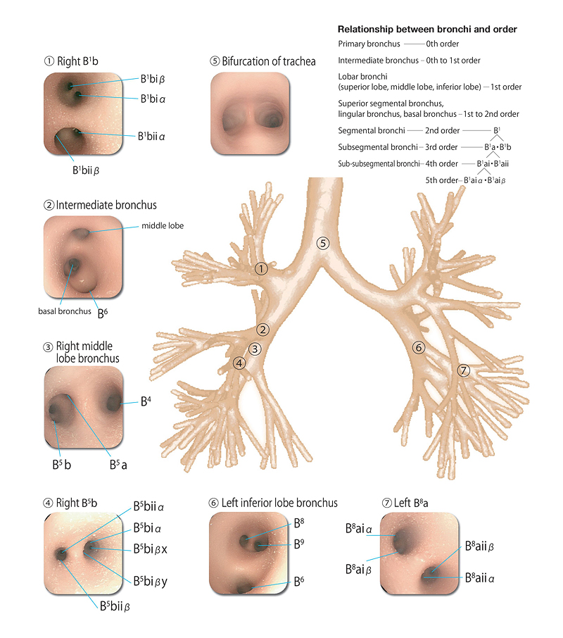
- The ultrathin structures of the bronchi in the bronchi main body were reproduced using a special manufacturing method, including the 5th order bronchi (e.g., B1aiα). Ultrathin bronchoscopes can be inserted up to the 5th order bronchi. ②
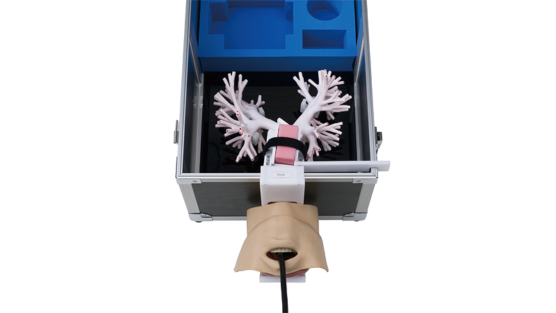
- A head model is attached, enabling insertion of the ultrasonic bronchoscope from the oral cavity and confirmation of the bifurcation of the bronchi and esophagus. ③
- The bronchi main body and the bronchi-support stand can be easily removed from the black case.
- The peripheral parts of the three-dimensional bronchi can be fully opened, enabling washing with water following endoscope insertion training.
- The material used is a special silicone rubber. Its elasticity confers a human body-like texture when inserting the bronchoscope. The inside of the bronchi is also similar in color to the human body.
Practical Training
- Endobronchial ultrasound-guided transbronchial needle aspiration (EBUS-TBNA) training
- External cylinder method training
- Ultrathin bronchoscopes insertion training up to the 5th order bronchi
- Confirmation of the bifurcation of the bronchi and esophagus
External cylinder method
This is such a method that as the external cylinder for puncture needle is pressed against the bronchial wall just before puncture, the tip of the external cylinder for the puncture needle is put into the recess between the cartilages to puncture by pushing the external cylinder together with a bronchoscope to forward and to backward. This method is expected to improve the diagnostic yield by securely collecting tissues while avoiding puncturing cartilages.
Takeo Inoue, Noriaki Kurimoto, et al.
New Technique for Endobronchial Ultrasound-guided Transbronchial Needle Aspiration to Improve Diagnostic Yield
J Bronchol Intervent Pulmonol 2013; 20: 28-32
Bronchoscopy image
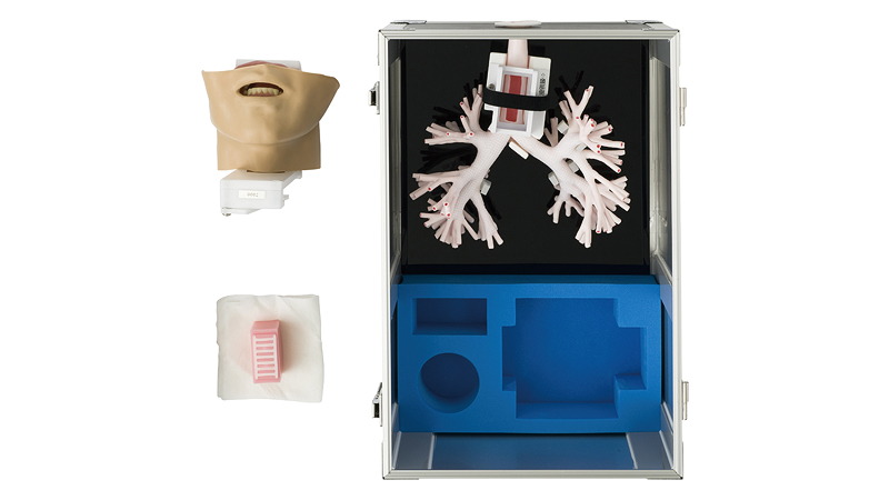
Components
 |
|
| Bronchi main body | 1 |
|---|---|
| Head model | 1 |
| Puncture site | 1 |
| Storage case (sponge case attached) | 1 |
Spares
| LM-099A | Puncture site (2 pcs) |
|---|---|
| LM-099B | Head model |
Specifications
Bronchus main body
| Size | Approx. 25(L) × 24(W) × 16(H) cm |
|---|---|
| Weight | Approx. 0.2 kg |
Storage case
| Size | Approx. 32(L) × 46(W) × 27(H) cm |
|---|---|
| Weight | Approx. 3.6 kg |
Head model
| Size | Approx. 17(L) × 16(W) × 15(H) cm |
|---|---|
| Weight | Approx. 0.7 kg |
Puncture site
| Size | Approx. 3(L) × 6(W) × 4(H) cm |
|---|---|
| Weight | Approx. 0.1 kg |