LM-106
KOKEN Tracheostomy Management Simulator LM-106


Outline
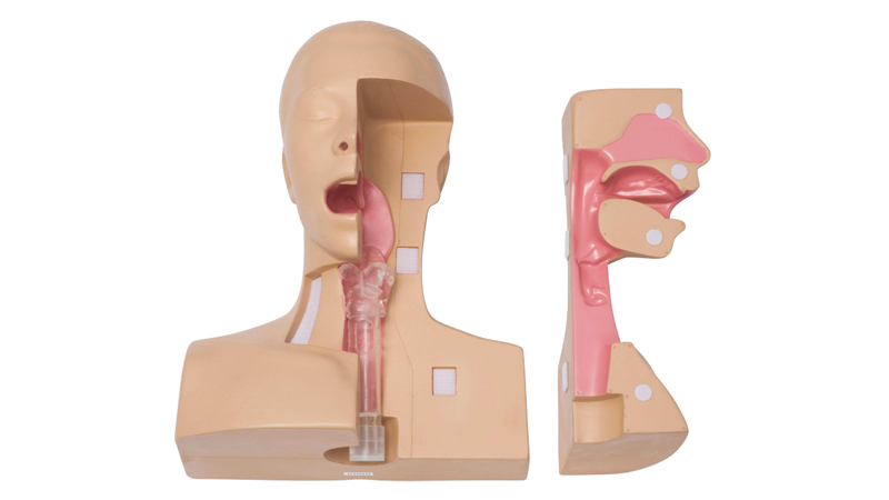
This model can be used to practice the procedures carried out for tracheostomy patients in the fields of nursing and caregiving when replacing cannulas or performing suction through a cannula.
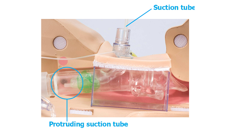
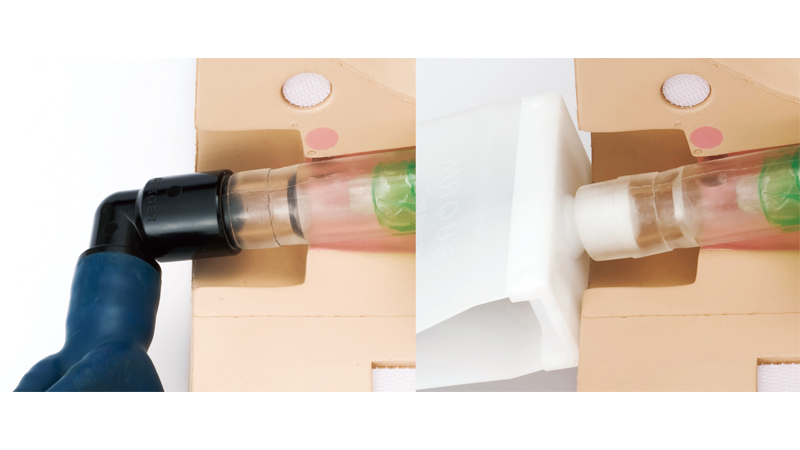
The model can be divided into two halves and the trachea section is transparent, allowing confirmation of the state of cannula insertion and the position of the suction catheter in the cannula during suction. In addition, with the specially provided cannula attached, a centilator can be operated by connection a test lung (simulated lung) to the model.
Features
- Allows the user to practice the procedures for replacing cannulas.
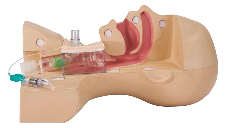
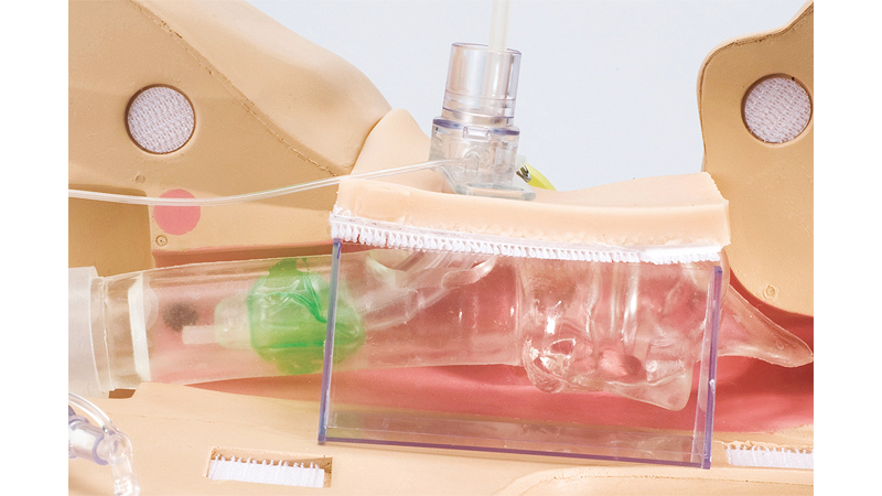
- As the trachea section is transparent, the state of inflation of the cuff can be observed, and by using the specially provided cannula, the approximate optimum pressure can be confirmed. (Because the posterior wall of the trachea is soft, the user can feel the compression of the trachea that results when the cuff is overinflated.)
- The transparent trachea facilitates explanation of how the suction catheter should be positioned, and of how to perform suction using the upper part of the cuff. (The above mentioned practicing of suction procedures can be performed using the simulated sputum.)
- The soft neck surface skin allows the user to feel the thyroid cartilage through the surface skin.
- The detachable trachea section facilitates explanation of the areas where granulation tends to develop.
- A ventilator can be operated by attaching the specially provided cannula and connecting a test lung, allowing confirmation of the alarm tone generated when air leakage occurs.
Practical Training
- As the trachea section is transparent, the state of inflation of the cuff can be observed, and by using the specially provided cannula, the approximate optimum pressure can be confirmed.
The detachable trachea section facilitates explanation of the areas where granulation tends to develop.
Example Use
- The transparent trachea facilitates explanation of how the suction catherter should be positioned, and of how to perform suction using the upper part of the cuff.
- A ventilator can be operated by attaching the specially provided cannula and connecting a test lung, allowing confirmation of the alarm tone generated when air leakage occurs.
Components
 |
|
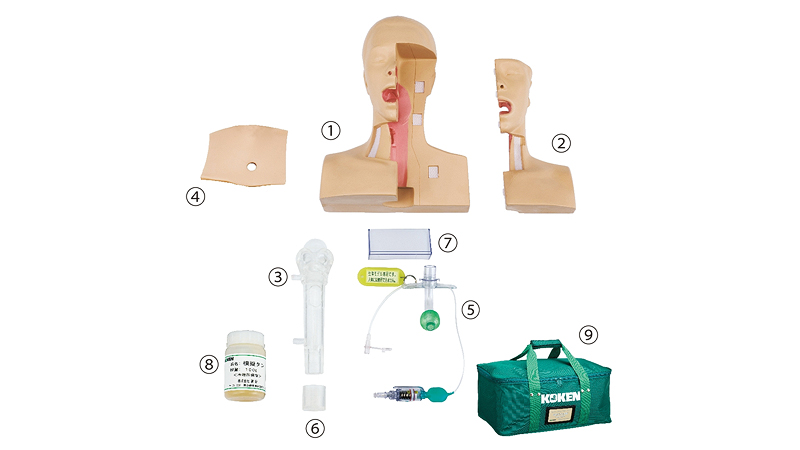
| ① Main body | 1 |
|---|---|
| ② Side of face | 1 |
| ③ Trachea | 1 |
| ④ Neck skin surface | 1 |
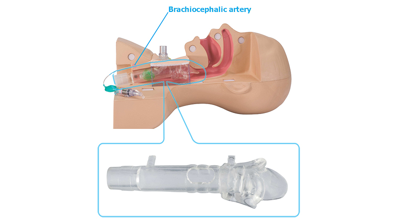
| ⑤ Specially provided tracheal cannula (The cuff is colored so that it can be identified when attached to the cannula) * Under no circumstances must this specially provided cannula be used on the living body |
1 |
| ⑥ Trachea cap | 1 |
| ⑦ Neck surface skin holding plate | 1 |
| ⑧ Simulated sputum (Viscosity can be adjusted diluting with water) | 1 |
| ⑨ Storage bag | 1 |
Spares
| LM-106A | Neck skin surface |
|---|---|
| LM-0701 | Simulated sputum (100g × 5pcs) |
Options
| LM-097A | Clasp for bronchus |
|---|---|
| LM-097D | Bronchus (with 2caps) |
* Bronchus for Suction Training Model Type Ⅱ can be attached to the trachea.
Specifications
| Size | Approx. 39(L) × 29(W) × 20(H) cm |
|---|---|
| Weight | Approx. 1.6 kg |