SonoScape P60

 ⇦ 

⇩ P60 Images ⇩  Home 

P60 new release in Medica

The timeless design of an ultrasound device is layered on a great foundation. Powered by the prominent Wis+ platform, P60 is a flagship ultrasound system offering exceptional image display and user-experience from the integration of state-of-the-art artificial intelligence technology. The AI functions now designed for holistic care for women: from family planning, pregnancy biometry and visualization to breast & pelvic health.

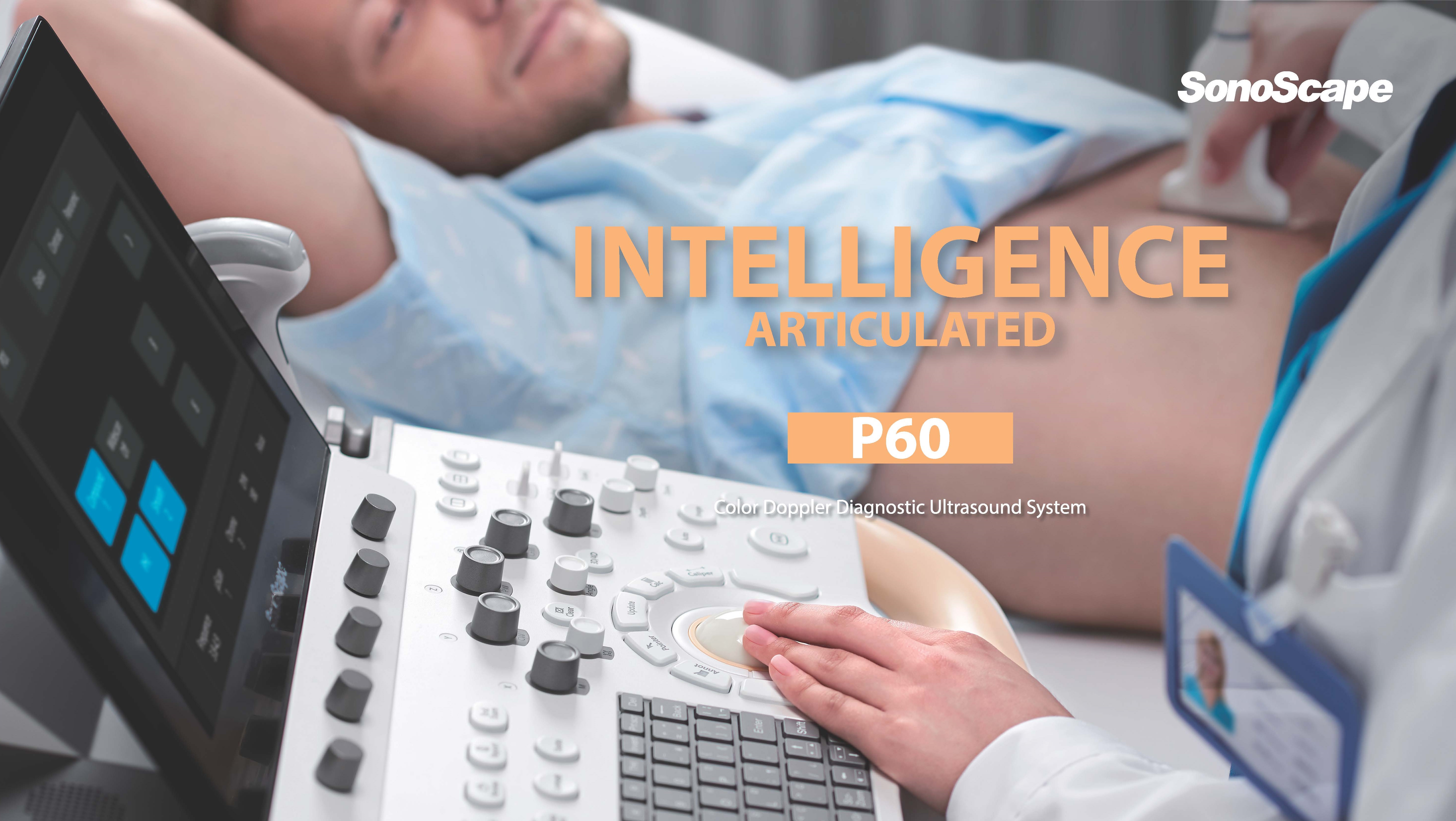
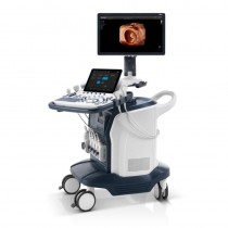
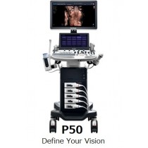
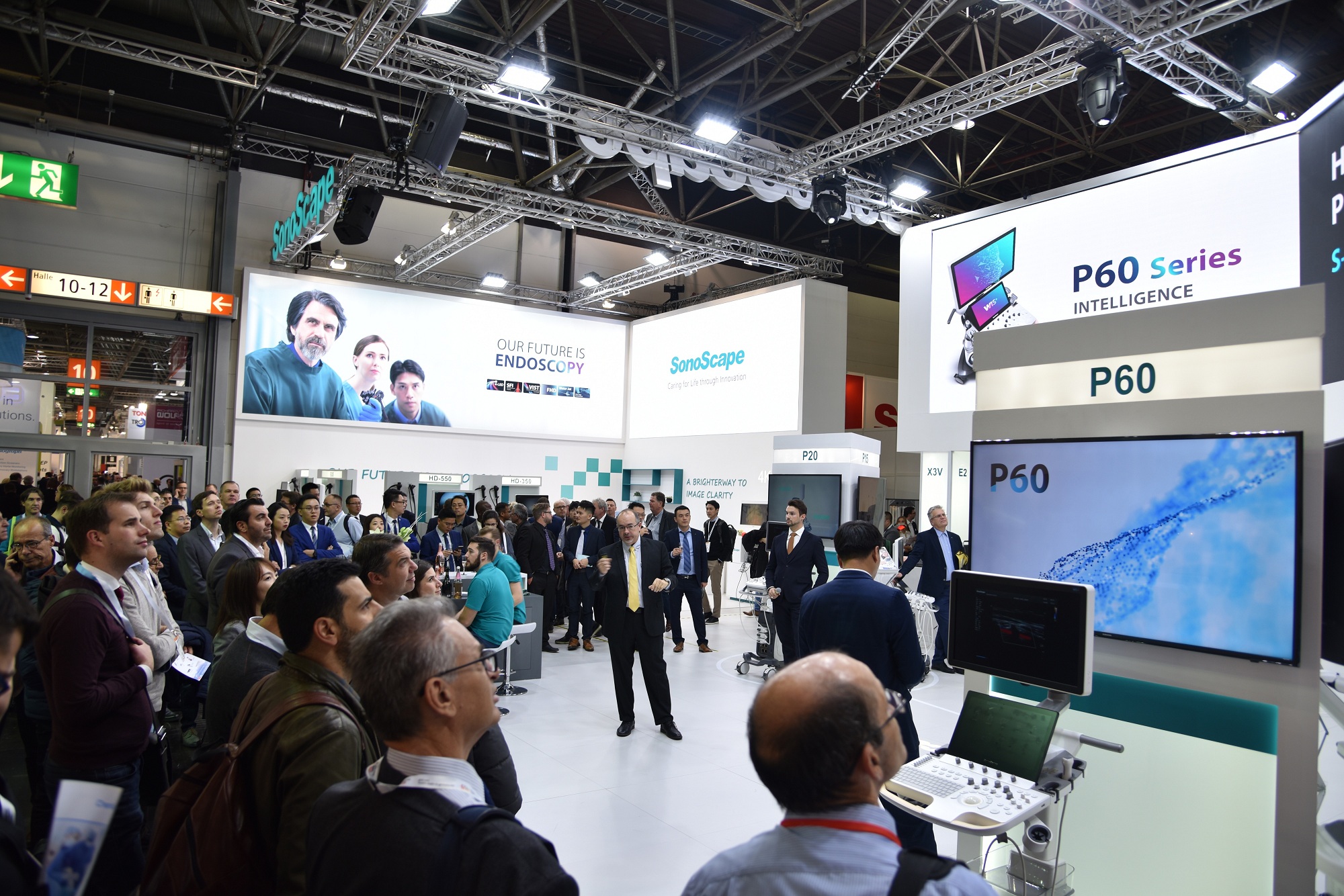
P60, configured with SonoScape's latest prominent Wis+ platform, is designed to provide more insightful and constructive evidence for diagnosis through authentic detail display, easy-but-effective intelligent analysis and streamlined workflow. Not only does P60 inherit SonoScape's consistent advantages in extraordinary imaging quality and optimized operation, but it also now benefits from the integration of state-of-the-art artificial intelligence technology and is dedicated to offering exceptional user-experience for a wide range of applications..

Compact yet Powerful Design

The design of P60 focues on simplicity and compactness but makes no compromise to powerful performance. Height adjustables and lateral rotatable panel and an articulating monitor arm can basically satisfy any requirements under different scanning conditions.

High Sensitivity Touch Panel User Friendly KeyBoard Power Assistant Battery Bulit-in Installed Gel Warmer

  • 24in LED Monitor(Option) 13.3in Tilting Touch Screen Built-in Battery
    P60 feature a large size 24inch high defination LED monitor,providing excellent image display to users. 13.3inch touch screen allows users to browse and select functions with wase. Atilting design works for adjustment in terms of users' needs.
    A built0in v\battery supports P60 to work for 2 hours without power suppoy, leaving users no worry sbout accidental pause and data loss due to power outage.
  • Consistent Quality Performance Across Applications
 ⇦  ⇩ CES Images  Home 

 Function of Micro 

Enables visualization for micro-vascularized structures

Micro F provides an innovative method to expand the range of visible flow in ultrasound, especially for visualizing slow flow tiny vessels. By adopting an advanced adaptive filter and accumulating temporal and spatial sognals, Micro F can distinguish minute flow from overlaying tissue movement effectively, and depict hemodynamic with higher sensitivitynand spaltial resolution. Detailed views of blood flow in relation to nearby tissue offered by Micro F render more diagnostic confidence to evaluate lesions and tumors.
  • Renal Anatomical structure Imaging

    Vascular visualiztion imaging

  • Abundant and Cervical Lymph Node Imaging

 ⇦  ⇩Cardiovascular Images Home 

 Function of CEUS 

Exerts the full potential of micro flow imaging

The comprehensive contrast-enhanced ultrasound imaging and quantification package on P60 offer doctors a through solution to evaluate perfusion dynamics in a wide range of clinical settings. Dynamic Acoustic control technology can generate a uniform acoustic pressure along the whole field and therefore elongate contrast agent duration and improve lesion perfusion. The combination of MFI, MFI Time and MFI Mix allows doctors to view the lesion perfusion from different perspectives and hence diagnose more easily and precisely.

  • Obstructive Cholestasis with MFI

    Micro Flow Imgaing automatically accumulates uptake of contrast agent and helps you trace small bubble populations, even in very low-perfused & peripheral areas.

  • HCC with Mix Mode

    Mix Mode offers an overlapped display of the contrast enhanced image together with the fundamental B mode image to help doctors better locate target lesion correctly.

  • HCC with MFI-Time

    MFI-Time helps to visualize both vascularization and perfusion intuitively by color coding the arrival times of contrast agents on different phases.

 ⇦  ⇩ Auto Button Function Home 

 Function of Advanced Cardiovascular 

Strives for a comprehensive solution for cardiac evaluation

The remarkable upgrade P60 has made on both imaging quality and accurate quantification makes it most versatile ever model of SonoScape in cardiovascular. Clear display of anatomical structures, hemodynamics and precise functional assessment are at the core of diagnosis evidence in cardiovascular imaging. Equipped with SonoScape's unique pure single crystal phased array transducers and state-of-the-art processing technology, P60 is committed to restore every fine detail and element for precise diagnosis. New Myocardium Quantitative Analysis(MQA) provides in-depth quantitative report on global and regional myocardial wall motion dynamics of the left ventricle, offering doctors a comprehensive assessment of myocardial functions.

  • New Myocardium Quantitaive (MQA)

    Extraordinary performance in 2D and color mode presents authentic cardiac anatomy details. Combianed with SonoScape's advanced wall motion tracking technology, P60 can provide precise analysis on myocardial functions.

  • Blood Flow

    Exquisite blood flow sensitivity, penetration, temporal and spatial resolution achieved by P60 clearly exhibits the profile of blood flow velocity and every detail of hemodynamics, even at deep parts.

 ⇦  ⇩ Artificial Intelligence Home 

 Function of Auto Button 


Auto Button

Auto is a shortcut key on the control panel that helps to adjust important imaging parameters automatically. It is available under B mode, CFM mode and PW mode. Moreover, it can be user-defined to activate AI-featured functions (S-Fetus, S-MSK, S-Breast, S-Thyroid), and therefore users won't be bothered searching on the touch screen. It is a unique design for saving doctors' much time and effort and allowing them to stay focused on the patient instead of being distracted with system operation.


Automated tools

Automated measurement and analysis tool package on P60 maker every exam more consistent, accurate and fast in different applications.

  • Auto EF

  • Auto IMT

  • Auto Blodder

  • Auto OB(NT)

 ⇦  ⇩ Intelligence Articulation Function Function Home 

 Function of Artificial Intelligence 

Brings unprecedented improvement on efficiency and accuracy

The adoption of AI on P60 not only simplifies the workflow greatly, but also provides enhanced reproducibility and consistency in measurement. With well-trained AI algorithms, the burdensome structure recognition and manual measurement procedures are now replaced with one-key operation. Given the big data used in the algorithms, the variables, which may affect the diagnosis conditions, could no more be a problem because of the uniform and precise guidelines built by AI. AI features are now available on S-Breast, S-Thyroid, S-Fetus and S-MSK.

S-Breast


  By simply setting ROI frame on a suspicious lesion, S-Breast helps to outline the lesion border and report the classification of suspicious breast lesions according to BI-RADS (Breast Imaging-Reporting and Data System) standard. The simplified workflow can both improve efficiency and provide standardized reporting on the classification of benign and malignant masses.

S-Thyroid


  S-Thyroid is an advanced tool in detecting and classifying suspicious thyroid lesions based on ACR TI-RADS(American College of Radiology Thyroid Imaging Reporting and Data System) guideline. After selecting the region of interest, S-Thyroid can automatically defines the lesion boundaries and generate a report regarding the features of the suspicious lesion.
 ⇦  ⇧ Top Home 

 Function of Intelligence Articulation 


 ⇦  ⇧ Top ⇧  Home 


  TOP


UltraSound Products ⊳ 

Request Demonstration 「html download pdf button icon」の画像検索結果

 ⇦ 


Home Product ⊳ 

Grid List

Set Ascending Direction

3 Item(s)

Grid List

Set Ascending Direction

3 Item(s)